
Quantitative Determination of Caspase-1 Expression in ATP and LPS Stimulated Monocytes by ELISA Analysis
Caspase family appears to be the key members involved in cellular apoptosis process and closely related with the morphological characteristics of apoptosis. It has been paid much attention by researchers during these years. Caspase-1 is the first discovered member, which was cloned and sequenced in 1992. After then, 14 Caspase members were cloned. Caspase-1, the key factor in the apoptosis mechanism, mainly involve in cytokine-mediated inflammatory response. Meanwhile, it plays a role in the death receptor-mediated cell apoptosis, taking part in the mature and transport of IL-1β, as well as related physiological and pathological process. During the stimulation of several factors such as growth factor, hit shock protein, cytokine and DNA damaging agent, it regulates several apoptosis transduction pathway.
The study on Caspase-1 expression is very popular in recent years. Among them, the hottest topic is stimulation of Caspase-1 by ATP and LPS. Fig.1. is the principle for ATP and LPS activation (Animal Biotechnology). When monocytes are stimulated with ATP and LPS, the expression of membrane receptor P2X7 increase, resulting in the influx of Ca2+ and outflow of K+. Expression of NLRP3 raises, then pro- Caspase-1 is activated and Caspase-1 increases.
The ELISA kit for Caspase-1 produced by Cloud-Clone Corp. can be used to determinate the concentration of Caspase-1 in THP-1 cells.
Experiment protocol:
1. Expression of Caspase-1 activated by ATP
(1) Collect human monocyte THP-1, adjust the concentration of cells to 106/ml using culture medium RPMI1640. Then cells are cultured in 5% incubator in 37℃ overnight.
(2) After incubation for 12 h, add LPS (1 ug/ml). Incubate for 3 h, then add ATP (5 mM) and incubate for 2 h.
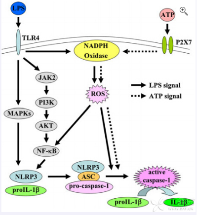
Fig.1. Signal pathway of Caspase-1 expression activated by ATP and LPS
2. Collection of culture supernates and lysates of TFP-1cells
Collect cultured cells. Centrifuge at 1000 g for 20 min, supernates are absorpted for later detection. Wash cell pellets with cold PBS for 3 times, lysis cells by ultrasonication or the freeze/thaw cycle. Centrifuge at 1000 g for 20 min and supernates are used for measurement.
3. Quantitative detection of concentration of Caspase-1 in THP-1 cells with Elisa kit SEB592Hu.
Results:
Fig.2. displays the concentration of Caspase-1 in cell supernates and lysates of THP-1 cells stimulated with ATP and LPS.
Discussion:
As showed in Fig.2., concentration of Caspase-1 in both THP-1 culture supernates and cell lysates increase after treated with ATP and LPS. Meanwhile, the increase is more significant in cell lysates. The concentration of Caspase-1 is about 24 times compared with untreated cell lysates, which demonstrates that Caspase-1 expresses within THP-1 cells.
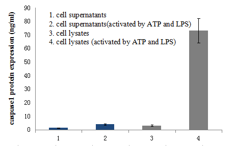
Fig.2. Caspase-1 expression in culture supernates and lysates after THP-1 cells were activated by ATP and LPS.