
Picture
Quality Guarantee

Certificate
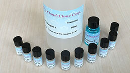
BCA Protein Quantification Kit
Instruction manual
First Edition (Revised on April, 2016)
PRODUCT INFORMATION
BCA Protein Assay Kit is based on one of the most popular protein concentration detection methods: BCA (bicinchoninic acid) method. The principle is the peptide bond of protein reacts with Cu2 + to form a complex, and Cu2 + is reduced to Cu + in alkaline environment. Then the BCA reagent binds with Cu + and forming stable colored complexes, which shows maximum absorbance at 562 nm. Consequently the protein content could be determined by the color of mixtures. It is fast, stable and sensitive, the lowest detection limit reached 20μg / ml, and it shows good linearity within 50-2000μg/ml.
REAGENTS AND MATERIALS PROVIDED
| Reagents | Size 1-100T | Size 2-500T | Size 3-2000T |
| BCA Reagent A | 20ml | 100ml | 400ml |
| BCA Reagent B | 1ml | 3ml | 12ml |
| BSA Standard | 2mg | 7.5mg | 30mg |
| Instruction manual | 1 | 1 | 1 |
STORAGE AND PERIOD OF VALIDITY
Store Reagent A and B at -4 ℃, and store BSA standards at -20 ℃. The Period of Validity is 1 year. NOTE: If sediments appeared in reagent A/B in valid time, please heat and stir gently to dissolve. If the kit changed color or been contaminated by microbes, please discard.
SAMPLE PREPARATION
1. Dilute or concentrate samples’ concentration to 20-2,000μg/ml.
2. Interference substances in samples can not be included or exceed the rated quantity.
3. The ways to remove interference substances in samples are shown below:
1) Dialysis or gel filtration;
2) By diluting, until the substances no longer interfere. It’s applicable only when the initial concentration is high, then it could fall in the detection range of quantitative analysis after dilution.
3)Precipitate proteins in samples with acetone or trichloroacetic acid (TCA). The liquid contains interfering substances could be discarded, and protein precipitation can be dissolved in ultrapure water, or alkali BCA working solution.
4) Increase the copperion’s content (mix the reagent A and B with 50: 2 or 50: 3 to get the working solution), which will eliminate the interference of copper chelator.
NOTE: The standards should be treated the same way with samples for high accuracy.
REAGENTS PREPARATION
1. Dilution of the Standards
Prepare a set of standards according to the table. Dilute the bovine serum albumin (BSA) in clean tubes, it’s better to use same buffer with the samples’. 1 bottle of 2mg/ml BSA is sufficient for standards in different detection range listed below, Each standard with specific concentration is enough for detection in triplicate .
Standard dilution schemes are listed as follows:
| Standard No. | Dilution Buffer Volume(μl) | Standard Reagent Volume(μl) | Final Concentration(μg/ml) |
| 1 | 0 | 300 | 2000 |
| 2 | 125 | 375μl of standard1 | 1500 |
| 3 | 325 | 325μl of standard 1 | 1000 |
| 4 | 175 | 175μl of standard 2 | 750 |
| 5 | 325 | 325μl of standard 3 | 500 |
| 6 | 325 | 325μl of standard 5 | 250 |
| 7 | 325 | 325μl of standard 6 | 125 |
| 8 | 400 | 100μl of standard 7 | 25 |
| 9 | 400 | 0 | 0 |
2. Add 50 fold Reagent A with 1 fold Reagent B (50: 1) to get the working solution, mix well,use it right after it was ready.
ASSAY PROCEDURES
1. Add 25μl of standards/samples to 96-well plates, respectively.
2. Add 200μl BCA working solution to each well, and shake gentlely to mix well, incubating at 37 ℃ for 30 minutes.
3. Take the plate out and cool to room temperature, then detect the absorbance at 562nm (If no 562nm, wavelength within 540nm-595nm is also suitable).
4. Fit the standard curve by the OD and relating concentration of standards and calculate the samples’ concentration (Quadratic equation fitting or 4 parameters fitting etc. are suggested. If the standard curve is drawn by hand, point drawing method is recommended.)
IMPORTANT NOTES
1. The detection range of the kit is 20-2,000μg/ml.
2. The content of each interference must be less than the critical value.
3. BCA working solution should be used right after it was ready.
TROUBLE SHOOTINGS
| Problems | Possible reasons | Solutions |
| No coloration of samples | Copper ion chelating agents existed | Dialysis, desalt or dilution. Increase the copperion’s content (mix the reagent A and B with 50: 2) |
| Low concentration in samples | Concentrate the samples | |
| The blank absorbance is normal, while standard and samples’ absorbance is low | The PH was changed by strong acid/alkali buffer | Dialysis, desalt or dilution |
| Wrong wavelength | Detect in 540nm-595nm | |
| High samples’ absorbance | The proteins’ concentration is high | Dilution |
| There’re lipids/lipoproteins in samples | Add 2%SDS to get rid of interferences on lipids. | |
| All wells including blank show deep purple | Reducing agents or amines contained (e.g., catecholamines) | Dialysis, or dilution |