p53 drives necroptosis via downregulation of sulfiredoxin and peroxiredoxin 3
On August 20, 2022, Juan Sastre, Department of Physiology, Faculty of Pharmacy, University of Valencia, Spain, and his team published a paper titled “p53 drives necroptosis via downregulation of sulfiredoxin and peroxiredoxin 3” in Redox Biology. They described a positive feedback between mitochondrial H2O2 and p53 that downregulates sulfiredoxin and peroxiredoxin 3 leading to necroptosis in inflammation and obesity.
The kits [ELISA Kit for High Mobility Group Protein 1 (HMGB1), SEA399Mu] of Cloud-Clone brand was chosed in this article, we are so proud for supporting the reaserchers.

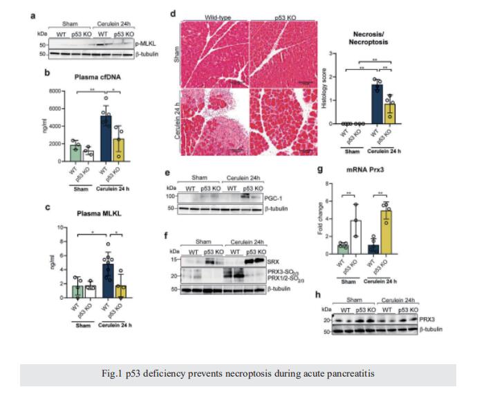
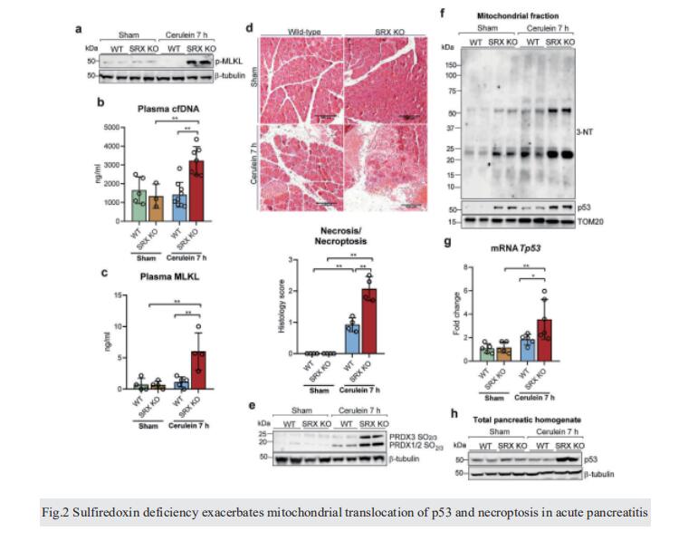
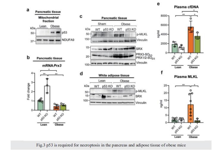
Mitochondrial dysfunction is a key contributor to necroptosis. We have investigated the contribution of p53, sulfiredoxin, and mitochondrial peroxiredoxin 3 to necroptosis in acute pancreatitis. Late during the course of pancreatitis, p53 was localized in mitochondria of pancreatic cells undergoing necroptosis. In mice lacking p53, necroptosis was absent, and levels of PGC-1α, peroxiredoxin 3 and sulfiredoxin were upregulated. During the early stage of pancreatitis, prior to necroptosis, sulfiredoxin was upregulated and localized into mitochondria. In mice lacking sulfiredoxin with pancreatitis, peroxiredoxin 3 was hyperoxidized, p53 localized in mitochondria, and necroptosis occurred faster; which was prevented by Mito-TEMPO. In obese mice, necroptosis occurred in pancreas and adipose tissue. The lack of p53 up-regulated sulfiredoxin and abrogated necroptosis in pancreas and adipose tissue from obese mice. We describe here a positive feedback between mitochondrial H2O2 and p53 that downregulates sulfiredoxin and peroxiredoxin 3 leading to necroptosis in inflammation and obesity.
