
Packages (Simulation)
Image (I)
-
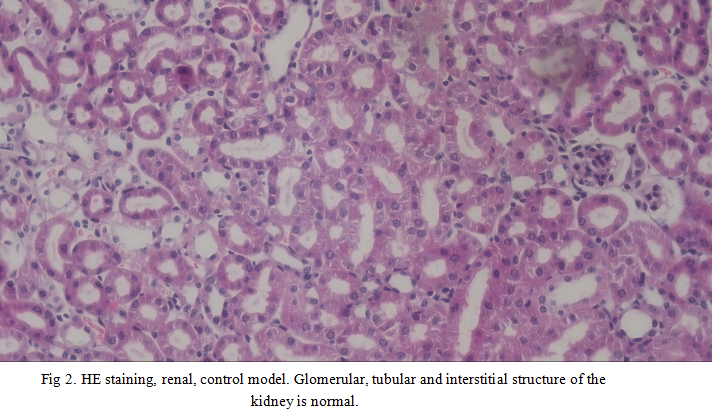 Fig 2. HE staining, renal, control model. Glomerular, tubular and interstitial structure of the kidney is normal.
-
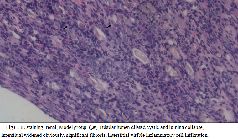 Fig3. HE staining, renal, Model group. (Arrow ) Tubular lumen dilated cystic and lumina collapse, interstitial widened obviously. significant fibrosis, interstitial visible inflammatory cell infiltration.
Image (II)
-
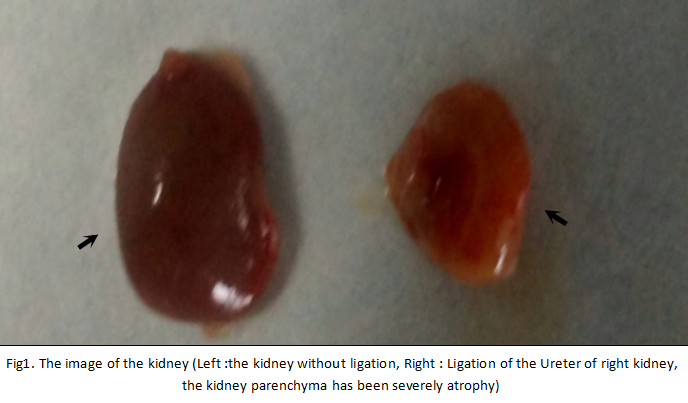 Fig 1.The image of the kidney (Left:withou ligation, Right:ligation)
Quality Guarantee

Certificate
Mouse Model for Renal Fibrosis (RF)
- Product No.DSI519Mu01
- Organism SpeciesMus musculus (Mouse) Same name, Different species.
- Prototype SpeciesHuman
- Sourceligation of unilateral ureter
- Model Animal StrainsBalb/c mice(SPF), healthy, male, week age:4W~6W, body weigh t:18g~20g
- Modeling GroupingRandomly divided into groups: Control group, Model group, Positive drug group and Test drug group, 15 mice per group.
- Modeling Period7d,14d,21d,28d
- Modeling Method1. 3% sodium pentobarbital (80mg/kg)intraperitoneal injection of anesthesia, remove the left back hair, skin disinfection.
2. Cut at 1cm of the left side of the spine on the top of the thigh, and cut the skin and muscles.
3. Separated the left ureter, dissociation of ureters at the 1/3 of the ureter and ligate twice with 6-0 at different site, and then cut ureter between the ligations, clean up the wound and make the two-layer suture. After the mice awake, put them back to clean cage, observe the state and death of mice, and make records.
4.kill the mice on 14d, 7d, 21d, 28d, respectively,take the left part of the kidney tissue, 4% poly formaldehyde solution fixed, paraffin embedded; the rest of the kidney tissue liquid nitrogen freezing and stored at -80℃. - ApplicationsDisease Model
- Downloadn/a
- UOM Each case
- FOB
US$ 180
For more details, please contact local distributors!
Model Evaluation
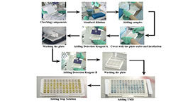
1.Urine protein and Urine NAG activity determination
Urinary protein and urinary NAG activity of the model group is significantly higher than that of the normal group and the sham operation group at 2 and 4 weeks after operation.
2. Serum creatinine (Scr), Urea nitrogen (BUN) content is determined after 4 weeks of operation, the model group serum Scr and BUN levels is increased.
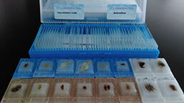
Pathological Results
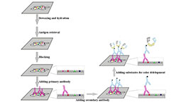
The left kidney is fixed with 4% poly formaldehyde, dehydrated, transparent, paraffin embedded and sliced. By HE staining and Masson staining, the pathological changes and inflammatory cell infiltration of glomerular and renal tubular interstitial are observed by light microscope.
In model group mice renal: interstitium inflammatory cell infiltration, renal tubular epithelial cell atrophy, loss of brush border, the tubular lumen expansion cystic, lumina collapse, interstitial widened obviously, interstitial visible inflammatory cell infiltration, accompanied by more into fibroblast proliferation. The inflammatory cell infiltration increased with time, the tubular epithelial cells were flat and detached, and the renal tubular atrophy, dilated, even collapsed, disappeared, collagen fiber hyperplasia, interstitial fibrosis increased.
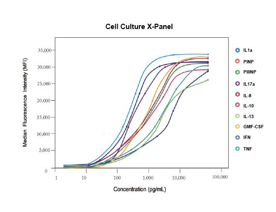
Cytokines Level
Statistical Analysis
SPSS software is used for statistical analysis, measurement data to mean ± standard deviation (x ±s), using t test and single factor analysis of variance for group comparison, P<0.05 indicates there was a significant difference, P<0.01 indicates there are very significant differences.
GIVEAWAYS
INCREMENT SERVICES
-
 Tissue/Sections Customized Service
-
 Serums Customized Service
-
 Immunohistochemistry (IHC) Experiment Service
-
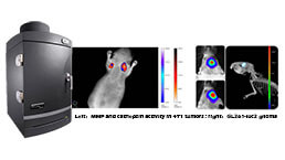 Small Animal In Vivo Imaging Experiment Service
-
 Small Animal Micro CT Imaging Experiment Service
-
 Small Animal MRI Imaging Experiment Service
-
 Small Animal Ultrasound Imaging Experiment Service
-
 Transmission Electron Microscopy (TEM) Experiment Service
-
 Scanning Electron Microscope (SEM) Experiment Service
-
 Learning and Memory Behavioral Experiment Service
-
 Anxiety and Depression Behavioral Experiment Service
-
 Drug Addiction Behavioral Experiment Service
-
 Pain Behavioral Experiment Service
-
 Neuropsychiatric Disorder Behavioral Experiment Service
-
 Fatigue Behavioral Experiment Service
-
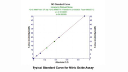
 Nitric Oxide Assay Kit (A012)
-
 Nitric Oxide Assay Kit (A013-2)
-
 Total Anti-Oxidative Capability Assay Kit(A015-2)
-
 Total Anti-Oxidative Capability Assay Kit (A015-1)
-
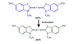
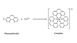
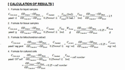
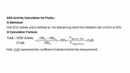 Superoxide Dismutase Assay Kit
-
 Fructose Assay Kit (A085)
-
 Citric Acid Assay Kit (A128 )
-
 Catalase Assay Kit
-
 Malondialdehyde Assay Kit
-
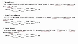
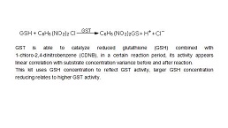 Glutathione S-Transferase Assay Kit
-
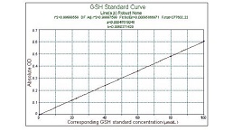
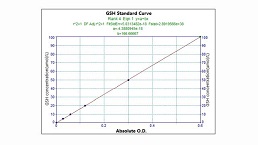 Microscale Reduced Glutathione assay kit
-
 Glutathione Reductase Activity Coefficient Assay Kit
-
 Angiotensin Converting Enzyme Kit
-
 Glutathione Peroxidase (GSH-PX) Assay Kit
-
 Cloud-Clone Multiplex assay kits
| Catalog No. | Related products for research use of Mus musculus (Mouse) Organism species | Applications (RESEARCH USE ONLY!) |
| DSI519Mu02 | Mouse Model for Renal Fibrosis (RF) | n/a |
| DSI519Mu03 | Mouse Model for Renal Fibrosis (RF) | n/a |
| DSI519Mu01 | Mouse Model for Renal Fibrosis (RF) | Disease Model |