
Packages (Simulation)
Image (II)
-
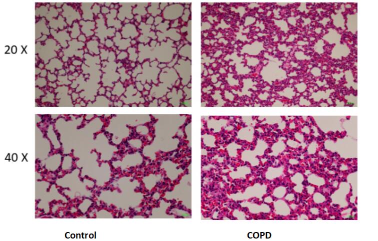 Fig. HE staining for lung
Quality Guarantee

Certificate


Mouse Model for Chronic Obstructive Pulmonary Disease (COPD)
- Product No.DSI557Mu02
- Organism SpeciesMus musculus (Mouse) Same name, Different species.
- Prototype SpeciesHuman
- Sourceinduced by smoking +LPS
- Model Animal StrainsICR Mice(SPF), healthy, male, age: 8~10weeks, body weight:30g~35g.
- Modeling GroupingRandomly divided into six group: Control group, Model group, Positive drug group and Test drug group.
- Modeling Period4-6 weeks
- Modeling MethodMain reagents: Hongjinlong cigarettes (produced by China Tobacco Hubei Industrial LLC, flue-cured tobacco, each cigarette containing 9mg tar, 12mg carbon monoxide), lipopolysaccharide (LPS, Sigma, USA), TNF-alpha and IL-18 ELISA kit (Cloud-Clone Corp).
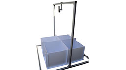
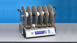
Modeling method: The SPF mice were fed for 1 week, and the animal model of COPD mice was established by intranasal infusion of LPS and smoking cigarettes. LPS (30μg/6μL) was instilled into the nasal cavity at 1st and 29th days, and from day 2 to 30 (except day 29), the mice were smoked and put into glass fumigator, 10 cigarettes per time, 30min per time, 5d per week, for 4 weeks in total. - ApplicationsDisease Model
- Downloadn/a
- UOM Each case
- FOB
US$ 200
For more details, please contact local distributors!
Model Evaluation
General observation: General condition Blank control group mice did not die, shiny hair, even breathing, normal activities; Mice in the model control group had dull hair, some hair removal phenomenon, restlessness, like to cluster when smoking, tired and curled up, sweating, abdominal distention, shortness of breath, nodding movement, even obvious mouth breathing.
Pathological Results
Morphological examination of lung tissue:
After abdominal anesthesia, the left lung was fixed in 10% formaldehyde solution for 24h, dehydrated paraffin embedded in conventional sections, stained with HE, and observed under light microscope.
In blank control group, the alveolar structure was clear, the alveolar size was uniform, the epithelial structure of airway mucosa was intact, and the cilia were arranged neatly.
In model control group, part of alveolar wall collapsed, alveoli expanded irregularly and fused with each other, forming bullae. Inflammatory cell infiltration of varying degrees could be seen in lung interstitium, bronchial mucosa epithelium exfoliated, mucosal folds increased, lumen protruded, cilia adhered and lumen narrowed, which was consistent with COPD pathological changes.
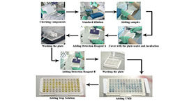
Cytokines Level
TNF Alpha test:
The mice were sacrificed within 24 hours after the last fumigation, and the mice were anesthetized. 1mL of blood was taken from the abdominal aorta of the mice, which was placed at room temperature for 2h, centrifuged at 2 000r/min for 20min, and the serum was taken and stored at -20℃ for use. The content of TNFalpha in serum of mice was detected according to the instructions of ELISA kit operation.
Statistical Analysis
SPSS software is used for statistical analysis, measurement data to mean ± standard deviation (x ±s), using t test and single factor analysis of variance for group comparison, P<0.05 indicates there was a significant difference, P<0.01 indicates there are very significant differences.
GIVEAWAYS
INCREMENT SERVICES
-
 Tissue/Sections Customized Service
-
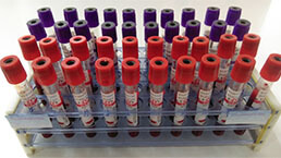 Serums Customized Service
-
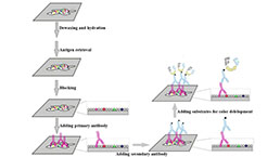 Immunohistochemistry (IHC) Experiment Service
-
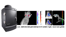 Small Animal In Vivo Imaging Experiment Service
-
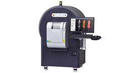 Small Animal Micro CT Imaging Experiment Service
-
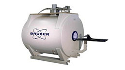 Small Animal MRI Imaging Experiment Service
-
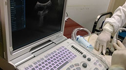 Small Animal Ultrasound Imaging Experiment Service
-
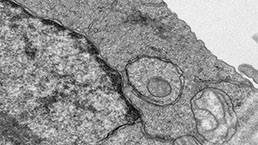 Transmission Electron Microscopy (TEM) Experiment Service
-
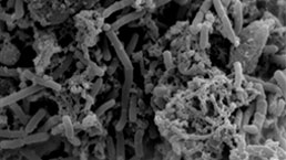 Scanning Electron Microscope (SEM) Experiment Service
-
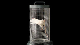
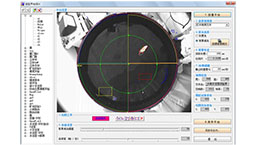 Learning and Memory Behavioral Experiment Service
-
 Anxiety and Depression Behavioral Experiment Service
-
 Drug Addiction Behavioral Experiment Service
-
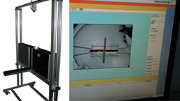
 Pain Behavioral Experiment Service
-
 Neuropsychiatric Disorder Behavioral Experiment Service
-
 Fatigue Behavioral Experiment Service
-
 Nitric Oxide Assay Kit (A012)
-
 Nitric Oxide Assay Kit (A013-2)
-
 Total Anti-Oxidative Capability Assay Kit(A015-2)
-
 Total Anti-Oxidative Capability Assay Kit (A015-1)
-
 Superoxide Dismutase Assay Kit
-
 Fructose Assay Kit (A085)
-
 Citric Acid Assay Kit (A128 )
-
 Catalase Assay Kit
-
 Malondialdehyde Assay Kit
-
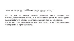 Glutathione S-Transferase Assay Kit
-
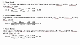
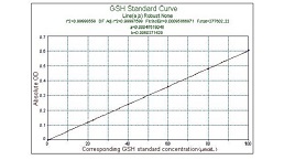
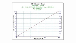 Microscale Reduced Glutathione assay kit
-
 Glutathione Reductase Activity Coefficient Assay Kit
-
 Angiotensin Converting Enzyme Kit
-
 Glutathione Peroxidase (GSH-PX) Assay Kit
-
 Cloud-Clone Multiplex assay kits
| Magazine | Citations |
| American Journal of Physiology. Endocrinology and Metabolism | Overexpression of histone deacetylase SIRT1 exerts an anti-angiogenic role in diabetic retinopathy via miR-20a elevation and YAP/HIF1α/VEGFA depletion Pubmed: 32776826 |
| ENVIRONMENT INTERNATIONAL | Circular RNA circBbs9 promotes PM2. 5-induced lung inflammation in mice via NLRP3 inflammasome activation Pubmed: 32707273 |
| Catalog No. | Related products for research use of Mus musculus (Mouse) Organism species | Applications (RESEARCH USE ONLY!) |
| DSI557Mu01 | Mouse Model for Chronic Obstructive Pulmonary Disease (COPD) | n/a |
| DSI557Mu02 | Mouse Model for Chronic Obstructive Pulmonary Disease (COPD) | Disease Model |
| TSI557Mu01 | Mouse Heart Tissue of Chronic Obstructive Pulmonary Disease (COPD) | Paraffin slides for pathologic research: IHC,IF and HE,Masson and other stainings |
| TSI557Mu03 | Mouse Liver Tissue of Chronic Obstructive Pulmonary Disease (COPD) | Paraffin slides for pathologic research: IHC,IF and HE,Masson and other stainings |
| TSI557Mu05 | Mouse Spleen Tissue of Chronic Obstructive Pulmonary Disease (COPD) | Paraffin slides for pathologic research: IHC,IF and HE,Masson and other stainings |
| TSI557Mu07 | Mouse Lung Tissue of Chronic Obstructive Pulmonary Disease (COPD) | Paraffin slides for pathologic research: IHC,IF and HE,Masson and other stainings |
| TSI557Mu09 | Mouse Kidney Tissue of Chronic Obstructive Pulmonary Disease (COPD) | Paraffin slides for pathologic research: IHC,IF and HE,Masson and other stainings |
| TSI557Mu15 | Mouse Brain Tissue of Chronic Obstructive Pulmonary Disease (COPD) | Paraffin slides for pathologic research: IHC,IF and HE,Masson and other stainings |
| TSI557Mu45 | Mouse Trachea Tissue of Chronic Obstructive Pulmonary Disease (COPD) | Paraffin slides for pathologic research: IHC,IF and HE,Masson and other stainings |
| TSI557Mu08 | Mouse Lung Tissue of Chronic Obstructive Pulmonary Disease (COPD) | Frozen tissues can be used for samples of Westerm Blot,Q-PCR,sequencing and protein,DNA extraction |