
Packages (Simulation)
Image (I)
-
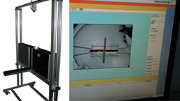
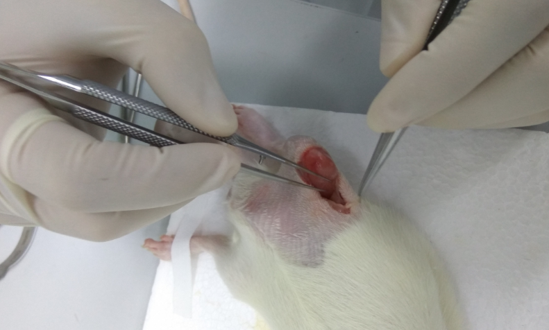 Fig: Fracture operation for rat leg
Image (II)
-
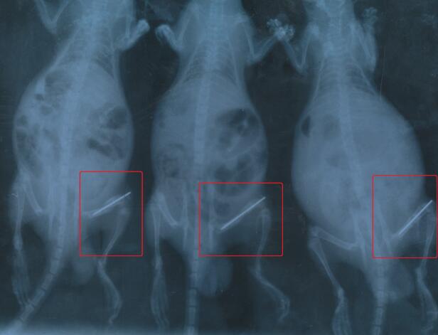 Fig: X-ray image of rat fracture legs
-
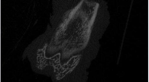 Fig. Micro CT imaging for rat femur
Quality Guarantee

Certificate

Rat Model for Fracture
Bone healing
- Product No.DSI797Ra01
- Organism SpeciesRattus norvegicus (Rat) Same name, Different species.
- Prototype SpeciesHuman
- SourceInduced by surgical method
- Model Animal StrainsSD rats (SPF class), Male, 6~8W, 200~250g
- Modeling GroupingRandomly divided into six group: Control group, Model group, Positive drug group and Test drug group
- Modeling Period6~8 weeks
- Modeling Method1. After intraperitoneal anesthesia in rats, the left lower limb of rats to wool, disinfection.
2. Make the tibial longitudinal incision in aseptic operation, cut the skin and muscle layer, exposed tibia in lower tibial 1/3 junction, with blades excluding 3mm wide periosteum, revealed subperiosteal bone, the bone rongeur snapped, causing a fracture.
3. Kirschner wire (0.8mm diameter ) were through the knee, straight into the fracture distal fracture, and then the fracture to be reset, the tail close to the top of the knee, and then layer by layer of muscle and skin, do not do fixed.
4. Intraperitoneal injection of penicillin 20U everyday for 3 days. - ApplicationsDisease Model
- Downloadn/a
- UOM Each case
- FOB
US$ 280
For more details, please contact local distributors!
Model Evaluation
1. X-ray photography: 1, 2, 3, 4, 5 weeks after surgery, take the right lower limb X-ray film.
2. X-ray examination for samples: anatomical fracture of the local callus tissue, the visual observation of fracture healing. Remove muscle and other soft tissue, femoral specimen removal of Kirschner wire, the use of X-ray machine shooting positive and lateral film.
3. Micro-CT examination: the specimen placed in the sample holder, adding 40% ethanol solution as a scanning medium, and then the sample holder placed in the 70KPa vacuum box for 20 minutes to ensure that no interference inside and outside the sample bubble. Select the scan parameters to scan the specimen.
Pathological Results
1. The callus tissue into the volume fraction of 4% paraformaldehyde fixed 24h, 4~6 weeks after decalcification, embedding, sectioning;
2. HE staining, Masson staining;
3. Immunohistochemical staining (IHC): ALP, BMP, PDGF, VEGF, Runx2, etc..
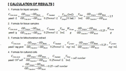
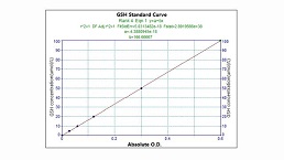
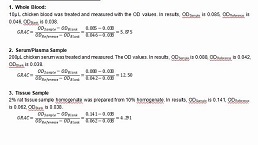
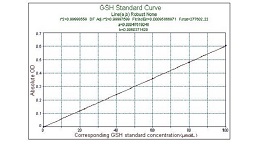
Cytokines Level
Statistical Analysis
SPSS software is used for statistical analysis, measurement data to mean ± standard deviation (x ±s), using t test and single factor analysis of variance for group comparison, P<0.05 indicates there was a significant difference, P<0.01 indicates there are very significant differences.
GIVEAWAYS
INCREMENT SERVICES
-
 Tissue/Sections Customized Service
-
 Serums Customized Service
-
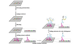 Immunohistochemistry (IHC) Experiment Service
-
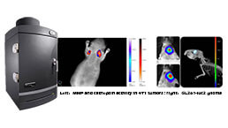 Small Animal In Vivo Imaging Experiment Service
-
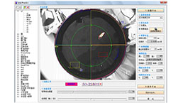
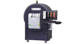 Small Animal Micro CT Imaging Experiment Service
-
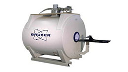 Small Animal MRI Imaging Experiment Service
-
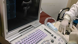 Small Animal Ultrasound Imaging Experiment Service
-
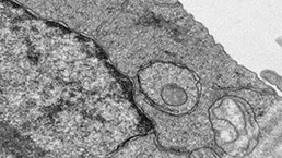 Transmission Electron Microscopy (TEM) Experiment Service
-
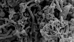 Scanning Electron Microscope (SEM) Experiment Service
-
 Learning and Memory Behavioral Experiment Service
-
 Anxiety and Depression Behavioral Experiment Service
-
 Drug Addiction Behavioral Experiment Service
-
 Pain Behavioral Experiment Service
-
 Neuropsychiatric Disorder Behavioral Experiment Service
-
 Fatigue Behavioral Experiment Service
-
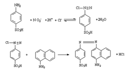 Nitric Oxide Assay Kit (A012)
-
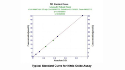 Nitric Oxide Assay Kit (A013-2)
-
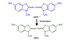 Total Anti-Oxidative Capability Assay Kit(A015-2)
-
 Total Anti-Oxidative Capability Assay Kit (A015-1)
-
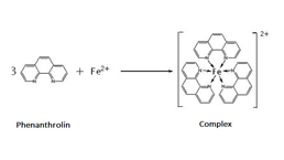
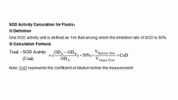 Superoxide Dismutase Assay Kit
-
 Fructose Assay Kit (A085)
-
 Citric Acid Assay Kit (A128 )
-
 Catalase Assay Kit
-
 Malondialdehyde Assay Kit
-
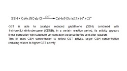 Glutathione S-Transferase Assay Kit
-
 Microscale Reduced Glutathione assay kit
-
 Glutathione Reductase Activity Coefficient Assay Kit
-
 Angiotensin Converting Enzyme Kit
-
 Glutathione Peroxidase (GSH-PX) Assay Kit
-
 Cloud-Clone Multiplex assay kits
| Catalog No. | Related products for research use of Rattus norvegicus (Rat) Organism species | Applications (RESEARCH USE ONLY!) |
| DSI797Ra01 | Rat Model for Fracture | Disease Model |