
Packages (Simulation)
Image (II)
-
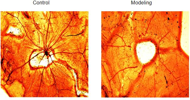 Fig. ADP staining of Retinal blood vessel
-
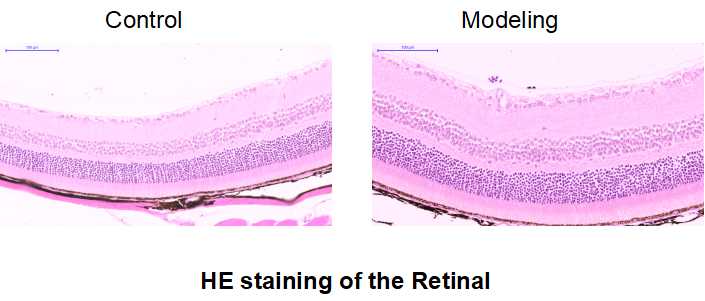 Fig. HE staining of the Retinal
Quality Guarantee

Certificate
Rat Model for Diabetic Retinopathy (DR)
DRP
- Product No.DSI841Ra01
- Organism SpeciesRattus norvegicus (Rat) Same name, Different species.
- Prototype SpeciesHuman
- SourceStreptozotocin (STZ) causes diabetes and induces retinopathy
- Model Animal StrainsSD Rats(SPF), healthy, male, bodyweight:180g~200g
- Modeling GroupingRandomly divided into six group: Control group, Model group, Positive drug group (Tiapride Hydrochloride )and Test drug group (three doses)
- Modeling Period8~10 weeks
- Modeling MethodModeling was conducted after 1 week of adaptive feeding. Fasting for 16h before diabetes model establishment, fasting weight was weighed, blood was collected from tail vein to measure blood glucose concentration, and streptozotocin STZ (65mg/kg) was injected intraderitoneal once. After 72h, 96h and 120h, random blood glucose was detected by steady glucose meter, body weight was measured, and general conditions such as body shape and hair color were observed. Tail venous blood was taken from each group to measure blood glucose. The diabetic mice with blood glucose ≥16.7mmol/L for three consecutive times and typical symptoms of "three more and one less" were identified as diabetic mice.
In healthy control group, STZ was changed to equal volume of normal saline, and other steps were the same.
Materials: 6 weeks after modeling, the left eye and right eye retinas were sacrificed.
The eyes were fixed with 4% paraformaldehyde for 24~48h. The sclera was cut open near the serrated edge of the ciliary body, the lens and vitreous were removed. The posterior segment of the eye was evenly divided into 3 parts, and the retina was gently float in water. Rinse with water for 2~3h. The retina was placed in a 3% trypsin digestion solution and incubated in a 37°C incubator for 2~3h. The retina was gently moved into distilled water and gently blown to completely remove the remaining retinal nerve components and inner boundary membrane, leaving a transparent layer of retinal vascular network. The retina was then moved to the loading plate to smooth as far as possible and dried naturally. - ApplicationsTo study the occurrence and treatment of diabetic retinopathy
- Downloadn/a
- UOM Each case
- FOB
US$ 300
For more details, please contact local distributors!
Model Evaluation
1. Retinal blood vessel staining
The cervical vertebrae of the model rats and the healthy mice in the control group were dislocated and sacrificed. The eyeballs were quickly removed and placed in PBS to carefully and quickly separate the retina. A radial incision was made with the optic papilla as the center, and the retina was tiled for ADP enzyme vascular staining.
In the control group, retinal blood vessels emanated from the optic disc, radially and evenly distributed in all directions, with thick diameter and good branches, and two layers of vascular network could be distinguished.
In the model group, there was no perfusion area in central retinal vessels, and blood vessels decreased and capillaries were lost.
Pathological Results
2. Retinal HE staining
The cervical vertebrae of model rats and control mice were dislocated and sacrificed. The eyeballs were quickly removed and fixed with 4% paraformaldehyde. After the fixation was completed, the ocular wall was cut at 0.5mm behind the tooth edge. The cornea, anterior segment and vitreous were carefully removed, and the retinal tissue was cut to prepare paraffin sections. HE staining was performed.
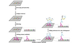
3. Immunohistochemical detection of retinal digestion tablet;
IHC detection was performed after the preparation of retinal patch.
4.QPCR detection: PI3K, AkT-1 mRNA levels
5.WB qualitative test: PTEN, BAD;
Cytokines Level
Statistical Analysis
SPSS software is used for statistical analysis, measurement data to mean ± standard deviation (x ±s), using t test and single factor analysis of variance for group comparison, P<0.05 indicates there was a significant difference, P<0.01 indicates there are very significant differences.
GIVEAWAYS
INCREMENT SERVICES
-
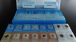 Tissue/Sections Customized Service
-
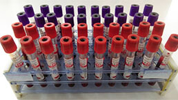 Serums Customized Service
-
 Immunohistochemistry (IHC) Experiment Service
-
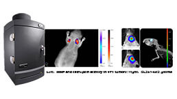 Small Animal In Vivo Imaging Experiment Service
-
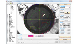
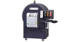 Small Animal Micro CT Imaging Experiment Service
-
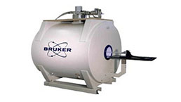 Small Animal MRI Imaging Experiment Service
-
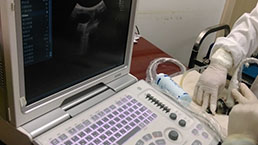 Small Animal Ultrasound Imaging Experiment Service
-
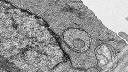 Transmission Electron Microscopy (TEM) Experiment Service
-
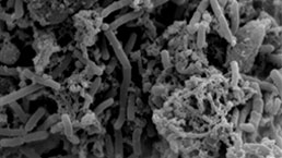 Scanning Electron Microscope (SEM) Experiment Service
-
 Learning and Memory Behavioral Experiment Service
-
 Anxiety and Depression Behavioral Experiment Service
-
 Drug Addiction Behavioral Experiment Service
-
 Pain Behavioral Experiment Service
-
 Neuropsychiatric Disorder Behavioral Experiment Service
-
 Fatigue Behavioral Experiment Service
-
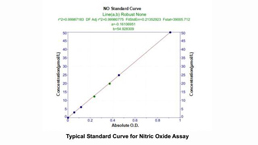
 Nitric Oxide Assay Kit (A012)
-
 Nitric Oxide Assay Kit (A013-2)
-
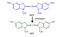 Total Anti-Oxidative Capability Assay Kit(A015-2)
-
 Total Anti-Oxidative Capability Assay Kit (A015-1)
-
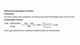 Superoxide Dismutase Assay Kit
-
 Fructose Assay Kit (A085)
-
 Citric Acid Assay Kit (A128 )
-
 Catalase Assay Kit
-
 Malondialdehyde Assay Kit
-
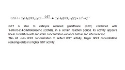 Glutathione S-Transferase Assay Kit
-
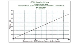
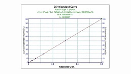 Microscale Reduced Glutathione assay kit
-
 Glutathione Reductase Activity Coefficient Assay Kit
-
 Angiotensin Converting Enzyme Kit
-
 Glutathione Peroxidase (GSH-PX) Assay Kit
-
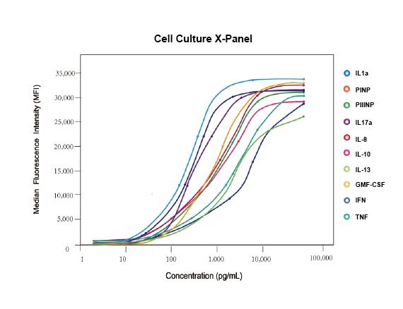 Cloud-Clone Multiplex assay kits
| Catalog No. | Related products for research use of Rattus norvegicus (Rat) Organism species | Applications (RESEARCH USE ONLY!) |
| DSI841Ra01 | Rat Model for Diabetic Retinopathy (DR) | To study the occurrence and treatment of diabetic retinopathy |