
Packages (Simulation)
Reagent Preparation
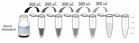
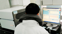
Image (I)
-
 Results demonstration
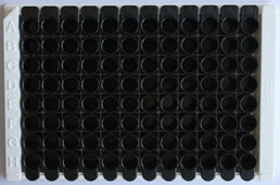
Image (II)
-
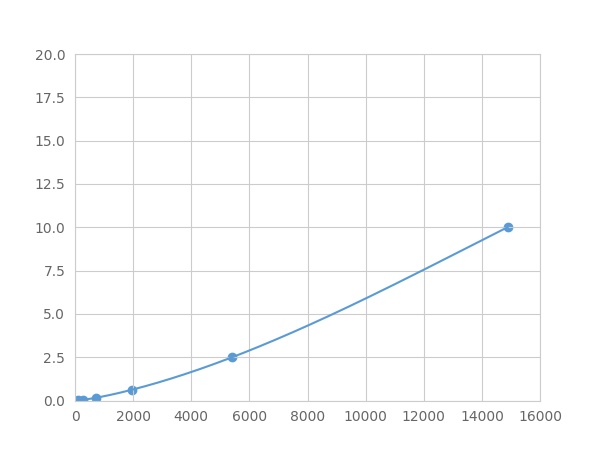 Typical Standard Curve
Quality Guarantee

Instruction Manual

Certificate


Multiplex Assay Kit for Peroxisome Proliferator Activated Receptor Gamma Coactivator 1 Alpha (PPARgC1a) ,etc. by FLIA (Flow Luminescence Immunoassay)
LEM6; PGC-1(alpha); PGC-1v; PGC1; PGC1A; PPARGC1; Ligand effect modulator 6
(Note: Up to 8-plex in one testing reaction)
- Product No.LMH337Hu
- Organism SpeciesHomo sapiens (Human) Same name, Different species.
- Sample TypeTissue homogenates, cell lysates and other biological fluids.
- Test MethodDouble-antibody Sandwich
- Assay Length3.5h
- Detection Range0.01-10ng/mL
- SensitivityThe minimum detectable dose of this kit is typically less than 0.003 ng/mL.
- DownloadInstruction Manual
- UOM 8Plex 7Plex 6Plex 5Plex 4Plex 3Plex 2Plex1Plex
- FOB
US$ 437
US$ 454
US$ 479
US$ 512
US$ 546
US$ 596
US$ 672
US$ 840
Add to Price Calculator
Result
For more details, please contact local distributors!
Specificity
This assay has high sensitivity and excellent specificity for detection of Peroxisome Proliferator Activated Receptor Gamma Coactivator 1 Alpha (PPARgC1a) ,etc. by FLIA (Flow Luminescence Immunoassay).
No significant cross-reactivity or interference between Peroxisome Proliferator Activated Receptor Gamma Coactivator 1 Alpha (PPARgC1a) ,etc. by FLIA (Flow Luminescence Immunoassay) and analogues was observed.
Recovery
Matrices listed below were spiked with certain level of recombinant Peroxisome Proliferator Activated Receptor Gamma Coactivator 1 Alpha (PPARgC1a) ,etc. by FLIA (Flow Luminescence Immunoassay) and the recovery rates were calculated by comparing the measured value to the expected amount of Peroxisome Proliferator Activated Receptor Gamma Coactivator 1 Alpha (PPARgC1a) ,etc. by FLIA (Flow Luminescence Immunoassay) in samples.
| Matrix | Recovery range (%) | Average(%) |
| serum(n=5) | 87-98 | 93 |
| EDTA plasma(n=5) | 79-91 | 81 |
| heparin plasma(n=5) | 93-101 | 97 |
| sodium citrate plasma(n=5) | 81-104 | 97 |
Precision
Intra-assay Precision (Precision within an assay): 3 samples with low, middle and high level Peroxisome Proliferator Activated Receptor Gamma Coactivator 1 Alpha (PPARgC1a) ,etc. by FLIA (Flow Luminescence Immunoassay) were tested 20 times on one plate, respectively.
Inter-assay Precision (Precision between assays): 3 samples with low, middle and high level Peroxisome Proliferator Activated Receptor Gamma Coactivator 1 Alpha (PPARgC1a) ,etc. by FLIA (Flow Luminescence Immunoassay) were tested on 3 different plates, 8 replicates in each plate.
CV(%) = SD/meanX100
Intra-Assay: CV<10%
Inter-Assay: CV<12%
Linearity
The linearity of the kit was assayed by testing samples spiked with appropriate concentration of Peroxisome Proliferator Activated Receptor Gamma Coactivator 1 Alpha (PPARgC1a) ,etc. by FLIA (Flow Luminescence Immunoassay) and their serial dilutions. The results were demonstrated by the percentage of calculated concentration to the expected.
| Sample | 1:2 | 1:4 | 1:8 | 1:16 |
| serum(n=5) | 80-88% | 88-95% | 91-101% | 85-105% |
| EDTA plasma(n=5) | 99-105% | 89-102% | 90-97% | 80-102% |
| heparin plasma(n=5) | 89-103% | 85-93% | 94-102% | 84-102% |
| sodium citrate plasma(n=5) | 86-97% | 78-101% | 96-103% | 93-101% |
Stability
The stability of kit is determined by the loss rate of activity. The loss rate of this kit is less than 5% within the expiration date under appropriate storage condition.
To minimize extra influence on the performance, operation procedures and lab conditions, especially room temperature, air humidity, incubator temperature should be strictly controlled. It is also strongly suggested that the whole assay is performed by the same operator from the beginning to the end.
Reagents and materials provided
| Reagents | Quantity | Reagents | Quantity |
| 96-well plate | 1 | Plate sealer for 96 wells | 4 |
| Pre-Mixed Standard | 2 | Standard Diluent | 1×20mL |
| Pre-Mixed Magnetic beads (22#:PPARgC1a) | 1 | Analysis buffer | 1×20mL |
| Pre-Mixed Detection Reagent A | 1×120μL | Assay Diluent A | 1×12mL |
| Detection Reagent B (PE-SA) | 1×120μL | Assay Diluent B | 1×12mL |
| Sheath Fluid | 1×10mL | Wash Buffer (30 × concentrate) | 1×20mL |
| Instruction manual | 1 |
Assay procedure summary
1. Preparation of standards, reagents and samples before the experiment;
2. Add 100μL standard or sample to each well,
add 10μL magnetic beads, and incubate 90min at 37°C on shaker;
3. Remove liquid on magnetic frame, add 100μL prepared Detection Reagent A. Incubate 60min at 37°C on shaker;
4. Wash plate on magnetic frame for three times;
5. Add 100μL prepared Detection Reagent B, and incubate 30 min at 37°C on shaker;
6. Wash plate on magnetic frame for three times;
7. Add 100μL sheath solution, swirl for 2 minutes, read on the machine.
GIVEAWAYS
INCREMENT SERVICES
| Magazine | Citations |
| Coronary Artery Disease | Impaired myocardium energetics associated with the risk for new-onset atrial fibrillation after isolated coronary artery bypass graft surgery Pubmed: 24463787 |
| Molecular and cellular biochemistry | Skeletal muscle mitochondrial dysfunction precedes right ventricular impairment in experimental pulmonary hypertension Pubmed: 23099843 |
| Diabetes. | Inorganic nitrate promotes the browning of white adipose tissue through the nitrate-nitrite-nitric oxide pathway Pubmed:25249574 |
| Psychoneuroendocrinology | Maternal stress predicts altered biogenesis and the profile of mitochondrial proteins in the frontal cortex and hippocampus of adult offspring rats PubMed: 26143539 |
| Biochemistry (Moscow) Supplement Series B: Biomedical Chemistry | The level of circulating PGC1α in cardiovascular diseases Article: 10.1134 |
| Neuroscience Letters | Unlike PPARgamma, neither other PPARs nor PGC-1alpha is elevated in the cerebrospinal fluid of patients with multiple sclerosis pubmed:28483651 |
| The American Journal of the Medical Sciences | Serum peroxisome proliferator-activated receptor gamma coactivator-1α related to myocardial energy expenditure in patients with chronic heart failure Doi: 10.1016/j.amjms.2018.12.002 |
| Brain Research | The effect of antecedent-conditioning high-intensity interval training on BDNF regulation through PGC-1α pathway following cerebral ischemia Pubmed: 31866362 |
| Food & Function | Ubiquinol supplementation modulates energy metabolism and bone turnover during high intensity exercise Pubmed: 32797125 |
| Brown and beige adipose tissue regulate systemic metabolism to resist diet-induced obesity through metabolite signals in an inter-organ signaling axis | |
| CLINICAL NUTRITION | Long lasting protective EFFECTS of early L-ARGININE TREAMENT on ENDOTHELIUM in an in Vitro STUDY 33743287 |
| Nature Communications | Brown and beige adipose tissue regulate systemic metabolism through a metabolite interorgan signaling axis 33772024 |