
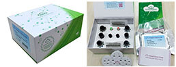
Packages (Simulation)
Reagent Preparation
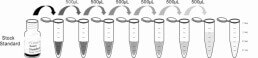
Image (I)
-
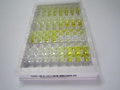 Results demonstration
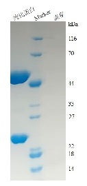
Image (II)
-
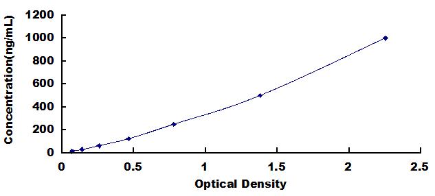 Typical Standard Curve
Quality Guarantee

Instruction Manual
Certificate


Maybe you can also pay attention to our smart microplate reader
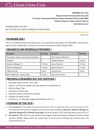
ELISA Kit for Lectin Galactoside Binding, Soluble 3 Binding Protein (LGALS3BP)
MAC-2-BP; LGALS3-BP; Galectin-3-Binding Protein; L3 Antigen; Mac-2-Binding Protein; Tumor-associated antigen 90K; Serum Protein 90K; Basement membrane autoantigen p105
- Product No.SEB766Hu
- Organism SpeciesHomo sapiens (Human) Same name, Different species.
- Sample Typeserum, plasma, tissue homogenates, cell lysates, cell culture supernates and other biological fluids
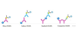
- Test MethodDouble-antibody Sandwich
- Assay Length3h
- Detection Range15.6-1,000ng/mL
- SensitivityThe minimum detectable dose of this kit is typically less than 6.1ng/mL.
- DownloadInstruction Manual
- UOM 48T96T 96T*5 96T*10 96T*100
- FOB
US$ 466
US$ 665
US$ 2993
US$ 5653
US$ 46550
For more details, please contact local distributors!
Specificity
This assay has high sensitivity and excellent specificity for detection of Lectin Galactoside Binding, Soluble 3 Binding Protein (LGALS3BP).
No significant cross-reactivity or interference between Lectin Galactoside Binding, Soluble 3 Binding Protein (LGALS3BP) and analogues was observed.
Recovery
Matrices listed below were spiked with certain level of recombinant Lectin Galactoside Binding, Soluble 3 Binding Protein (LGALS3BP) and the recovery rates were calculated by comparing the measured value to the expected amount of Lectin Galactoside Binding, Soluble 3 Binding Protein (LGALS3BP) in samples.
| Matrix | Recovery range (%) | Average(%) |
| serum(n=5) | 83-94 | 87 |
| EDTA plasma(n=5) | 78-105 | 101 |
| heparin plasma(n=5) | 83-99 | 91 |
Precision
Intra-assay Precision (Precision within an assay): 3 samples with low, middle and high level Lectin Galactoside Binding, Soluble 3 Binding Protein (LGALS3BP) were tested 20 times on one plate, respectively.
Inter-assay Precision (Precision between assays): 3 samples with low, middle and high level Lectin Galactoside Binding, Soluble 3 Binding Protein (LGALS3BP) were tested on 3 different plates, 8 replicates in each plate.
CV(%) = SD/meanX100
Intra-Assay: CV<10%
Inter-Assay: CV<12%
Linearity
The linearity of the kit was assayed by testing samples spiked with appropriate concentration of Lectin Galactoside Binding, Soluble 3 Binding Protein (LGALS3BP) and their serial dilutions. The results were demonstrated by the percentage of calculated concentration to the expected.
| Sample | 1:2 | 1:4 | 1:8 | 1:16 |
| serum(n=5) | 98-105% | 85-98% | 89-103% | 91-98% |
| EDTA plasma(n=5) | 83-99% | 78-102% | 98-105% | 78-92% |
| heparin plasma(n=5) | 97-105% | 80-101% | 79-91% | 79-90% |
Stability
The stability of kit is determined by the loss rate of activity. The loss rate of this kit is less than 5% within the expiration date under appropriate storage condition.
To minimize extra influence on the performance, operation procedures and lab conditions, especially room temperature, air humidity, incubator temperature should be strictly controlled. It is also strongly suggested that the whole assay is performed by the same operator from the beginning to the end.
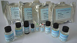
Reagents and materials provided
| Reagents | Quantity | Reagents | Quantity |
| Pre-coated, ready to use 96-well strip plate | 1 | Plate sealer for 96 wells | 4 |
| Standard | 2 | Standard Diluent | 1×20mL |
| Detection Reagent A | 1×120µL | Assay Diluent A | 1×12mL |
| Detection Reagent B | 1×120µL | Assay Diluent B | 1×12mL |
| TMB Substrate | 1×9mL | Stop Solution | 1×6mL |
| Wash Buffer (30 × concentrate) | 1×20mL | Instruction manual | 1 |
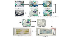
Assay procedure summary
1. Prepare all reagents, samples and standards;
2. Add 100µL standard or sample to each well. Incubate 1 hours at 37°C;
3. Aspirate and add 100µL prepared Detection Reagent A. Incubate 1 hour at 37°C;
4. Aspirate and wash 3 times;
5. Add 100µL prepared Detection Reagent B. Incubate 30 minutes at 37°C;
6. Aspirate and wash 5 times;
7. Add 90µL Substrate Solution. Incubate 10-20 minutes at 37°C;
8. Add 50µL Stop Solution. Read at 450nm immediately.
GIVEAWAYS
INCREMENT SERVICES
-
 Single-component Reagents of Assay Kit
-
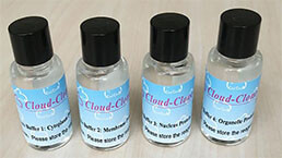 Lysis Buffer Specific for ELISA / CLIA
-
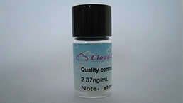 Quality Control of Kit
-
 ELISA Kit Customized Service
-
 Disease Model Customized Service
-
 Serums Customized Service
-
 TGFB1 Activation Reagent
-
 Real Time PCR Experimental Service
-
 Streptavidin
-
 Fast blue Protein Stain solution
-
 Single-component Reagents of FLIA Kit
-
 Streptavidin-Agarose Beads
| Magazine | Citations |
| Andrology | Comparative proteomic analysis coupled with conventional protein assay as a strategy to identify predictors of successful testicular sperm extraction in patients with non-obstructive azoospermia PubMed: 23427166 |
| Liver International | Identification of candidate biomarkers for hepatocellular carcinoma in plasma of HCV-infected cirrhotic patients by 4-D DIGE Pubmed: 23944848 |
| Acta Anaesthesiologica Scandinavica | Early prognostic factors in septic shock cancer patients: a prospective study with a proteomic approach Pubmed:29315472 |